
Dr. Marc Kaloustian
View ProductCase 5
miniKUT EZP was used at 800 rpm directly after access cavity preparation and orifice location. The working length was established. miniKUT OS 15/07 at 800 rpm was then used to relocate the canals in the coronal third prior to the final shaping. miniKUT 25/05 was finally used at 800 rpm, until the apical foramen and the corresponding gutta-percha cone was adapted to the canal and cone fit performed. Canals were filled with the continuous wave technique and PlanB Bioceramic Sealer.
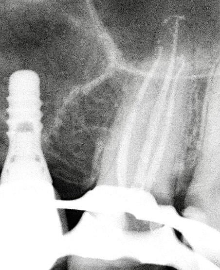
Case 4:
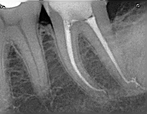
Case of a lower second left molar with an abrupt curvature in the distal canal. Canals were shaped with the miniKUT small canals pack procedure. The Glide path was directly performed with miniKUT 15/03 at 800 rpm in the 2 mesial canals. After preflaring with miniKUT OS file at 1000 rpm, the last 2 mm of the distal canals were scouted using a 08 hand file pre-bended with the EndoBender2 at 90 degrees. The deep shape was done with miniKUT 25/03 at 800 rpm in the mesial canals and in the 2/3 of the distal canal. The last 2 mm of the distal canals were only instrumented with 90 degrees pre-bent miniKUT 15/03 file introduced manually after many attempts. Canals were filled with a bioceramic sealer and continuous wave technique till mid root. Final xrays shows the respect of the anatomy in the apical part without canal transportation.
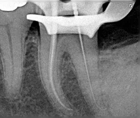
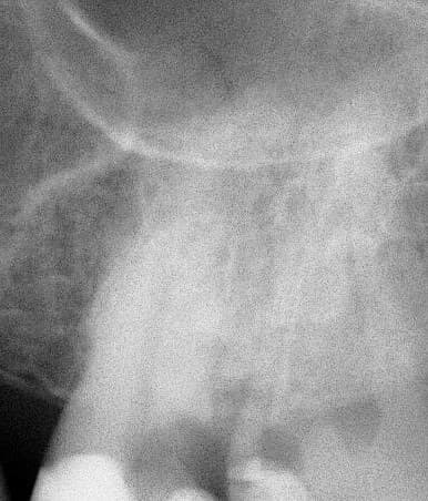
Case 3:
Case of an upper second molar with curved mesial canals. Canals were shaped with the miniKUT small canals pack procedure. The Glide path was directly performed with miniKUT 15/03 at 800 rpm in all 4 canals. MB1 and MB2 were confluent in the middle third. Deep shape was done with miniKUT 25/03 at 800 rpm. Canals were filled with a bioceramic sealer and continuous wave technique till mid root. Final xrays shows conservative shaping with respect of the original anatomy of the tooth.
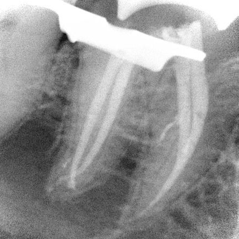
Case 2:
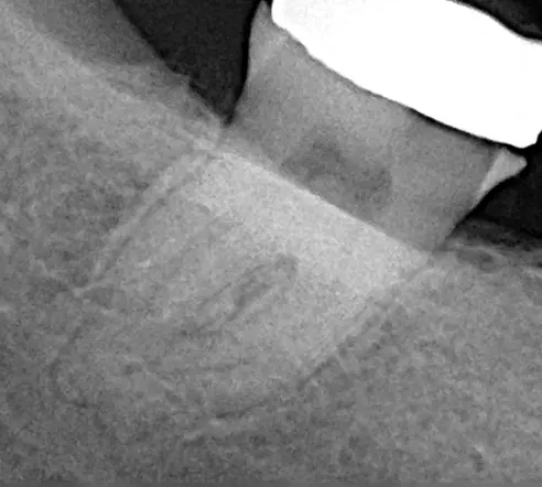
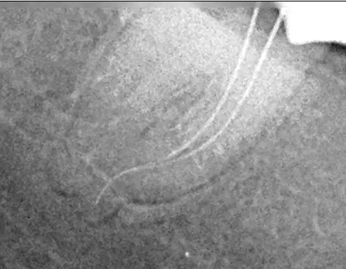
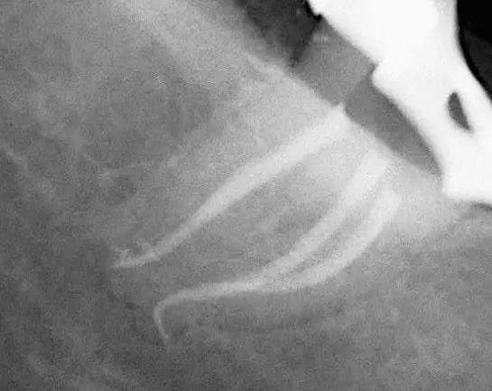
This case demonstrates using the miniKUT EZP 15/03 as “First File to Length”. The second image features no patency with 10K File. The third image shows patency with miniKUT 15/03 run at 800rpm.
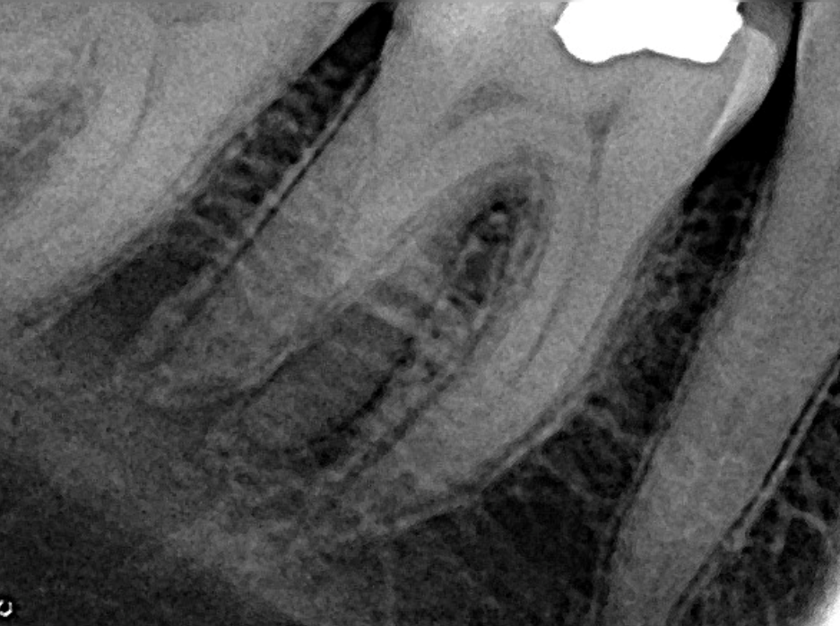
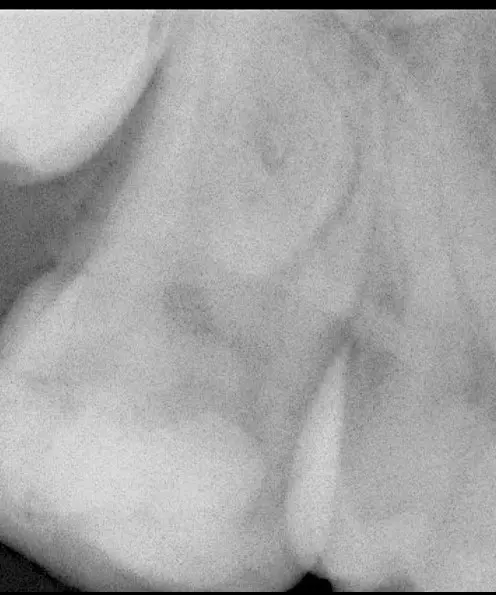
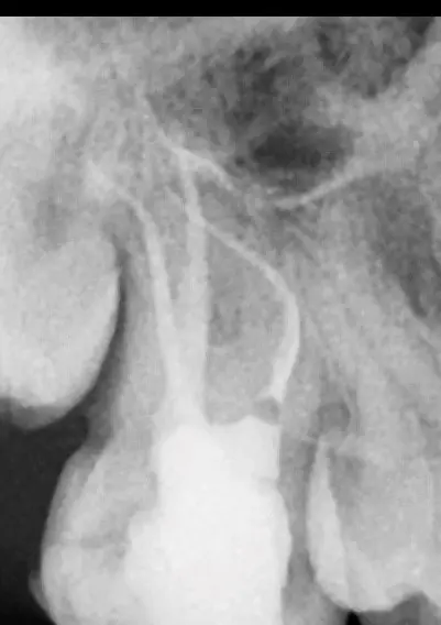
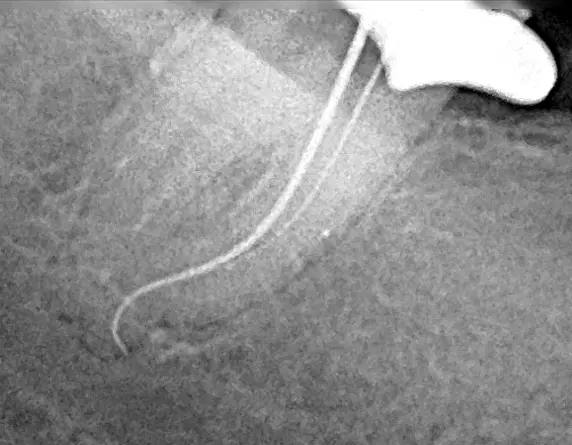
Case 1:
miniKUT EZP was used at 800 rpm directly without any prior use of handfiles after decay removal, access cavity preparation and orifice location. The working length was established with a 31 mm number 10 K file. miniKUT OS 15/07 at 800 rpm was then used to relocate the canals in the coronal third prior to the final shaping. miniKUT 25/05 was finally used at 800 rpm, until the apical foramen and the corresponding gutta-percha cone was adapted to the canal and cone fit performed. Canals were filled with the continuous wave technique.
Treatment of an upper second left molar with a deeply distal decay and pulp exposure. Patient was suffering from spontaneous pain increased at night. A diagnosis of irreversible pulpitis was established and a root canal treatment was performed in a single session. All canals were challenging because they were very narrow and long (23 to 25 mm) especially the mesio-buccal and disto-buccal canals. The disto buccal canal had also a small apical abrupt curvature.